Standards regarding the number of dental photos
Dentysta stomatolog Warszawa Ursynów ADENTIS / Information and advice / Dental advice / Standards regarding the number of dental photos

ACTION X-ray diagnostics – for health
This post is dedicated to all those who are healthy and may they always be healthy.
It is worth performing X-ray diagnostics
A dentist is a DOCTOR who needs precise tools to prepare a good treatment plan.
My dears, please DO NOT be afraid of having an X-ray done at the dentist.
Please be concerned when this test is NOT performed.
The doctor orders the test when it is needed.
X-ray diagnostics in detecting malocclusion in CHILDREN and ADULTS, in dental treatment, in prosthetic treatment, in implantological treatment is necessary.
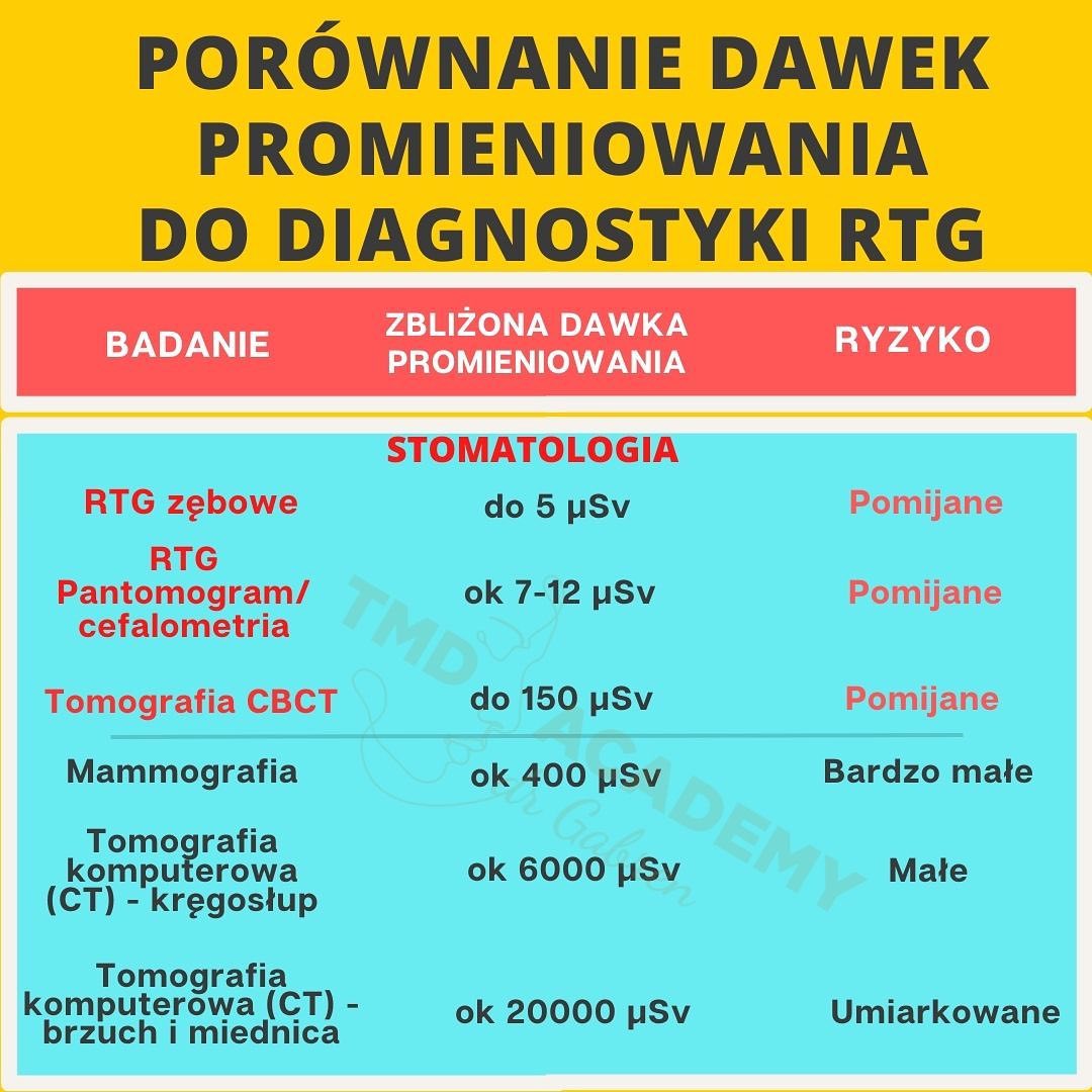
For example, the dose of radiation that we absorb during one photo is lower than the dose to which we are exposed during a flight from Krakow to the USA. It corresponds to approximately 3 hours of flight. The radiation from a digital tomograph (CBCT) is negligible compared to the radiation reaching the Earth from space.
We are constantly exposed to radiation.
Ps. Precise diagnostics can improve your health, sometimes it can save your life.
I ordered cbct for a patient and detected a pre-cancerous lesion in the sinus. The patient has already finished the procedure. The surgeon could have acted at the right time. Everything ended with SUCCESS.
Trust your DENTIST
Pursuant to the Regulation of the Council of Ministers of January 18, 2005 on limit doses of ionizing radiation (Journal of Laws of February 3, 2005), the limit dose for members of the general population is 1 mSv during a calendar year, while for eye lenses it is 15 mSv, and for the skin 50 mSv for any area of 1 cm2 of the irradiated part of the skin. This dose may be exceeded in a given calendar year, provided that in the following five calendar years its total value does not exceed 5 mSv.
There are no guidelines on how many photos or X-rays can be taken at one time. In radiological diagnostics, the basic principle of radiological protection ALARA (As Low As Reasonably Achieveable) should be followed. This means that as many exposures should be performed as are necessary to provide new information in the diagnostic process that will influence the method and outcome of treatment. However, you should avoid taking photos that are not important for achieving the diagnostic goal and therefore do not influence the choice of treatment method and, consequently, its result. The legal basis in this case is the Regulation of the Minister of Health of August 25, 2005 on the conditions for the safe use of ionizing radiation for all types of medical exposure (Journal of Laws 05.194.1625), Art. 33c § 4 point 1 says: “Diagnostic tests using ionizing radiation are performed in a way that guarantees the achievement of the required result with the lowest possible radiation dose.” That’s why at ADENTIS we only take radiological images in digital form.
It is worth knowing that the effective dose for the patient when taking an intraoral photo on film with sensitivity E is approximately 1.6 µSv, i.e. only 0.16% of the annual dose limit. In the case ofdigital photos, the effective dose is several times lower, depending on the imaging system used. Taking 7 conventional dental photos is approximately 11.2 µSv, which is less than half of the exposure dose when taking a panoramic X-ray and one tenth of the dose received when taking a chest X-ray. As a reminder, the dental status is the 20 intraoral photos taken most often at the same time (16 dental and 4 bitewing), which in many countries (e.g. in the USA) is used to visualize the teeth instead of a panoramic X-ray.
Dr Aleksandra Gabren